Left Acetabular Roof Mri

Acetabular Angle Radiology Reference Article Radiopaedia Org

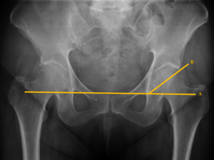
This Radiograph Documents The Measurement Of The Acetabular Roof Download Scientific Diagram

Stress Fracture In Acetabular Roof Due To Motocross Case Report

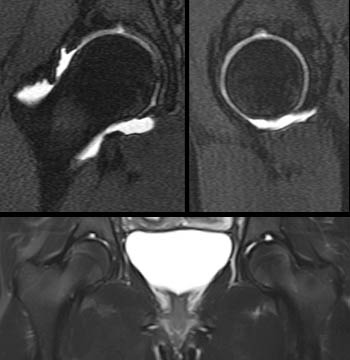
Representative Example In Borderline Dysplastic Hip A Coronal Mri Download Scientific Diagram

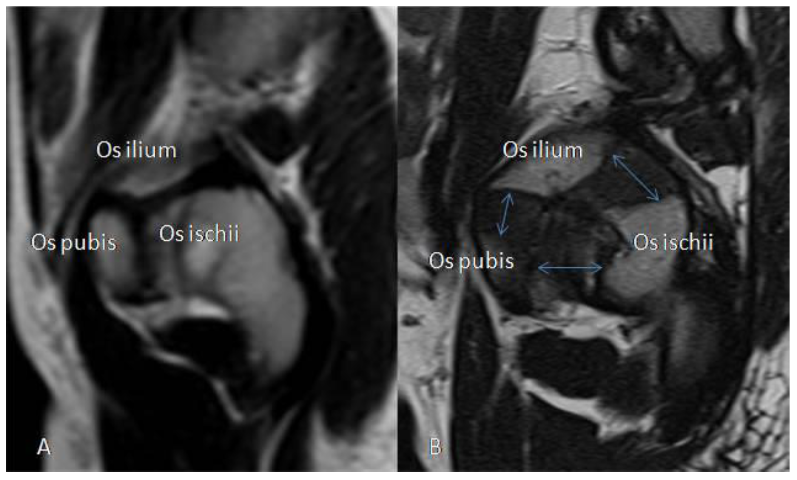
Mri Anatomy Of The Hip Review Mri Anatomy Of The Hip
Http Links Lww Com Jbjs E539

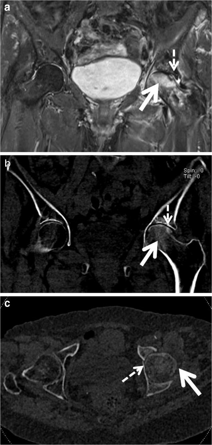
Metastatic lesions localized in the periacetabular area cause troublesome pain and reduced mobility of the patients.
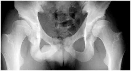
Left acetabular roof mri.
Understanding Clinical Radiology Of Fracture Acetabulum Trauma International
A Case Of Growth Disturbance Of The Acetabular Roof 5 3 Years Following Download Scientific Diagram
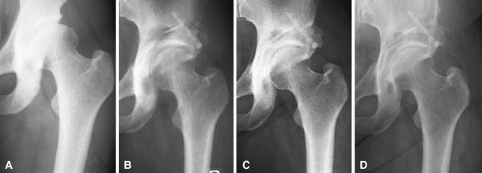
Developmental Dysplasia Of The Hip Radiology Reference Article Radiopaedia Org

Sketch Of The Three Main Types Of Acetabular Roof Dysmorphia A Download Scientific Diagram
Hip Disorders Radiology Key

Ct A And Mri T2w Fat Sat B At 2 Months After Surgery The Ct Scan Download Scientific Diagram

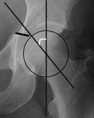
Measurement Of The Acetabular Index Ai From The Lateral End Of The Download Scientific Diagram
Os Acetabuli Radiology Reference Article Radiopaedia Org
Presentation1 Radiological Imaging Of Developmental Dysplasia Of The

Stress Fracture In Acetabular Roof Due To Motocross Case Report Sciencedirect

A Plain Radiograph Showing A Left Displaced Acetabular Anterior Wall Download Scientific Diagram

Imaging Of The Hip A Systematic Approach To The Young Adult Hip Abstract Europe Pmc

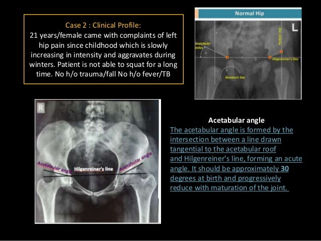
Girl With Left Hip Pain Mechanical Symptoms

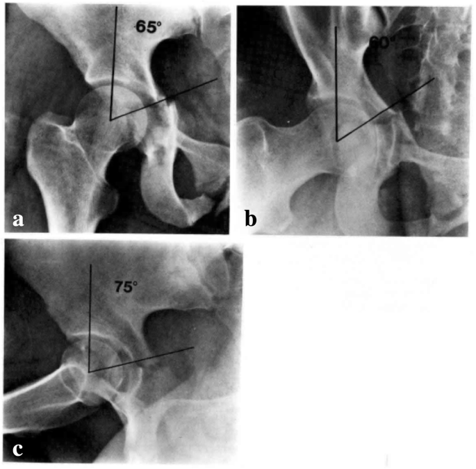
A The Center Edge Angle Of Wiberg Dce Acetabular Roof Obliquity Download Scientific Diagram

Sclerotic Bone Leison Note Sclerotic Bone Leison In Tibia In Young Age Osteoid Osteoma Note Malignancy In Old Age Radiology Tumor Oral Pathology

Chapter 14 Pelvic Trauma Musculoskeletal Key

Radiographic Anatomy And Imaging Of The Acetabulum Sciencedirect
Http Pdf Posterng Netkey At Download Index Php Module Get Pdf By Id Poster Id 117296
Https Encrypted Tbn0 Gstatic Com Images Q Tbn 3aand9gctomex0gykvkjjeykmbpzcq5w25ug9wffu Trvyunwurzjyd4vm Usqp Cau

Pin By Contessa La Lobo On Peds Measuring Angles Radiology Ultrasound
Orif Through Modified Stoppa Approach For Anterior Column

Figure 2 From Desmoid Tumor Of Ilio Acetabular Region With Articular Cartilage Breach A Case Report Semantic Scholar

Pelvis And Perineum Radiology Key
Pelvic High Grade Chondrosarcoma Delayed Development Can Bisphosphonates Be Responsible A Case Report
Epos
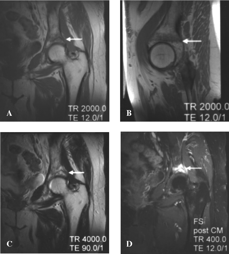
Figure 2 From Isolated Supra Acetabular Insufficiency Fracture A Case Report Semantic Scholar

Acetabular Fracture Radiology Reference Article Radiopaedia Org
Outcomes After Surgical Treatment Of Acetabular Fractures A Review Springerlink
Rotational Acetabular Osteotomy For Pre And Early Osteoarthritis Secondary To Dysplasia Provides Durable Results At 20 Years Springerlink

Trident Acetabulum Radiology Reference Article Radiopaedia Org

Pediatric Radiology

Angles Describing The Orientation Of The Acetabulum And Femoral Download Scientific Diagram
Femoroacetabular Impingement Radsource
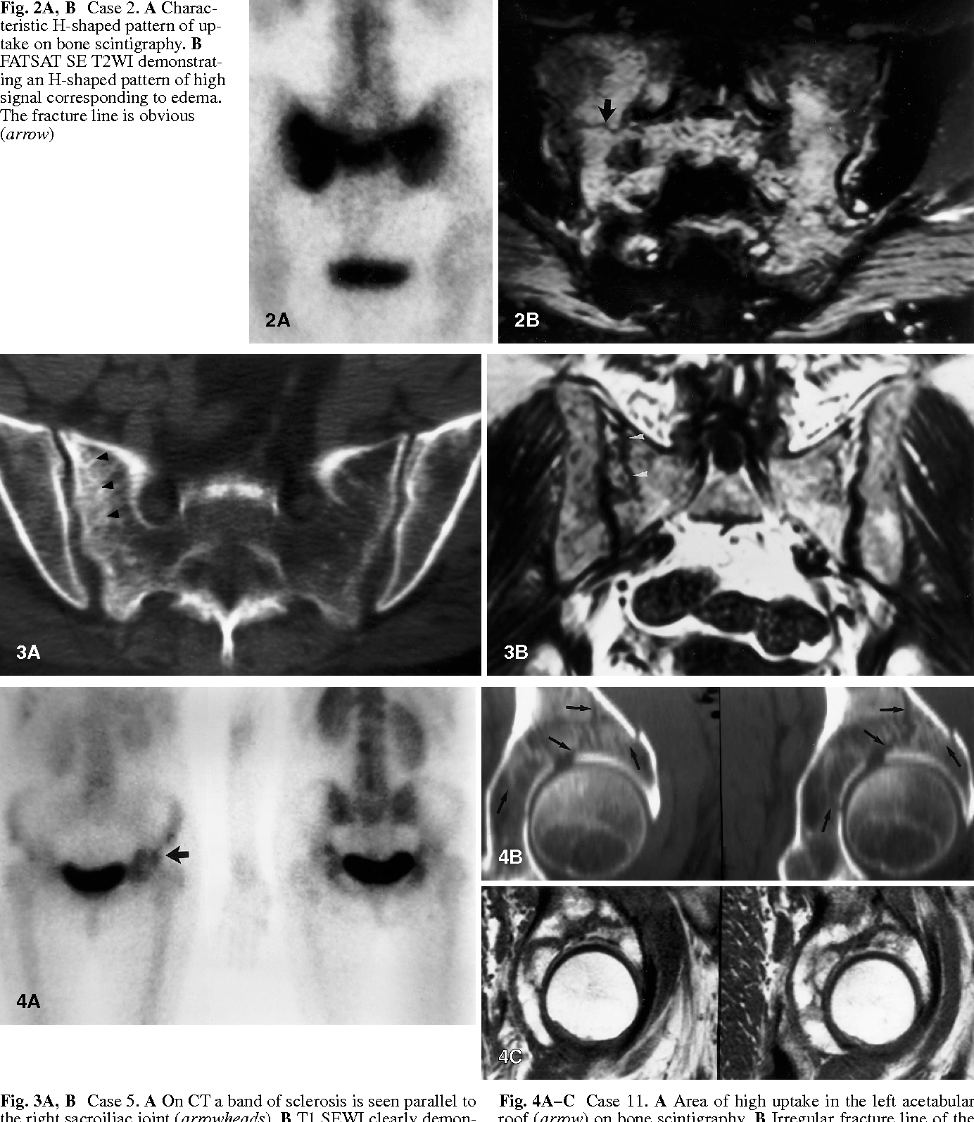
Role Of Mri In The Diagnosis Of Insufficiency Fractures Of The Sacrum And Acetabular Roof Semantic Scholar
Jcm Free Full Text Hip Morphology In Mucolipidosis Type Ii Html

Comprehensive Clinical Evaluation Of Femoroacetabular Impingement Part 2 Plain Radiography Sciencedirect

Epos Trade
Ddh Final Dt2
Bone Marrow Edema Around The Hip In Non Traumatic Pain Dual Energy Ct Vs Mri Springerlink
Imaging Of The Hip Radiology Key
Hip Dysplasia Information Symptoms Diagnosis Treatment

Congenital Disorders Of Bone And Joint Development Musculoskeletal Key
Acetabular Fractures
Https Encrypted Tbn0 Gstatic Com Images Q Tbn 3aand9gcq2jd7l23kq2utnhxntl9kdo8nlhr9lhqeohz6 Rr9vehqcxl42 Usqp Cau
Source : pinterest.com
