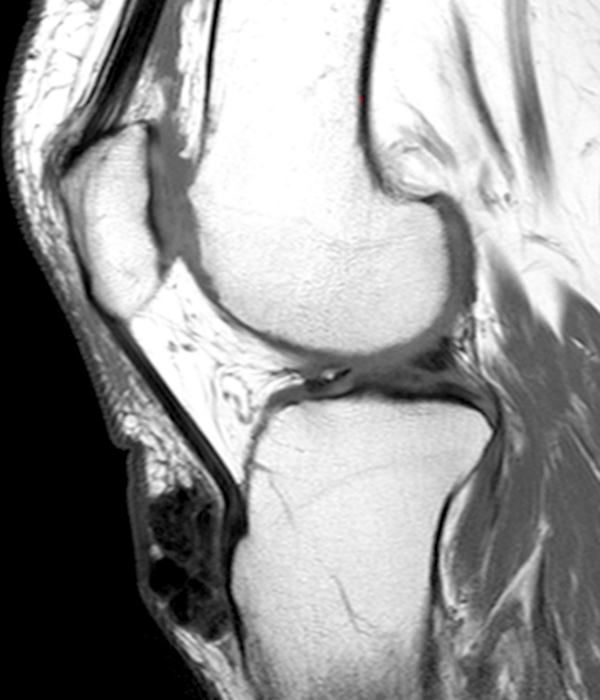
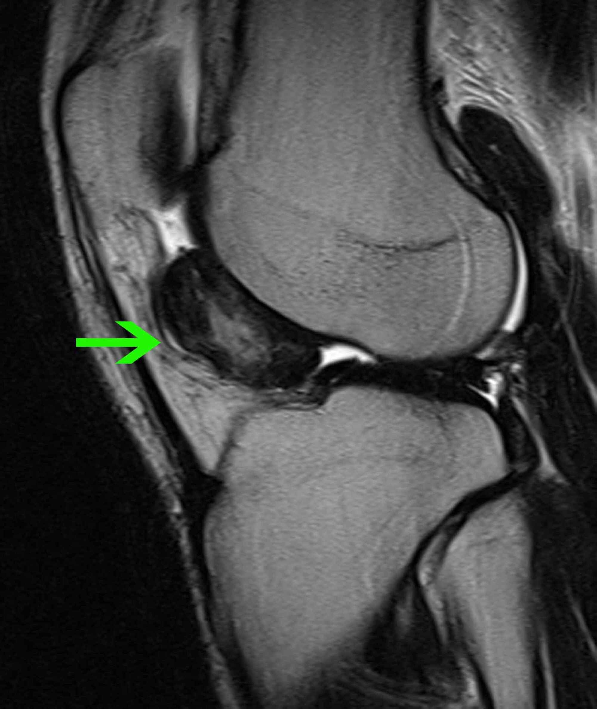
Lateral Gutter Knee Mri
Knee Bursae Radsource

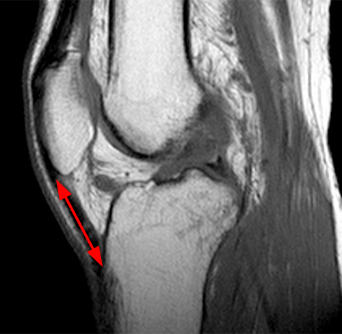
Mcl Tear On Coronal Mri Of Knee Magnetic Resonance Imaging Knee Mri Magnetic Resonance

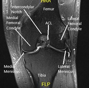
Mri Anatomy Of The Knee Ortopedia Y Traumatologia Anatomia Rodillas
Knee Mri Radiology Key
Arthrofibrosis Of The Knee Radsource

Mri Knee Anatomy Knee Sagittal Anatomy Free Cross Sectional Anatomy Knee Mri Mri Anatomy

Diagnostic arthroscopy is a crucial skill for diagnosing intra articular disorders of the knee including meniscal synovial ligamentous and articular cartilage pathology.
Lateral gutter knee mri.
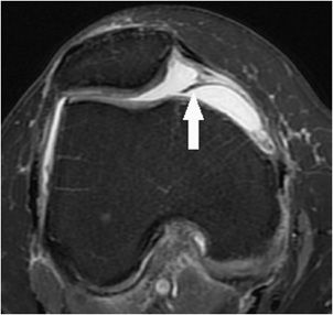
Synovial Plicae Of The Knee Radsource

Mri Knee Google Search Mri Anatomy Images Knee Mri

Mri Knee Acl Google Search Mri Knee Mri Radiology
Cureus Arthroscopic Technique For The Treatment Of Localized Pigmented Villonodular Synovitis Of The Knee
Case Study Management Of Ramp Lesion Of The Medial Meniscus With Acl Deficient Left Knee In A 30 Year Old Female

Mri Knee Anatomy Knee Mri Mri Anatomy Of The Knee

Bucket Handle Medial Meniscus Tear Radiology Case Radiopaedia Org Knee Mri Meniscus Tear Medial Meniscus Tear

Pin On Anatomy Imaging

Double Pcl Sign Appears On Sagittal Mri Images Of The Knee When A Bucket Handle Tear Of A Meniscus Most Commonly The Me Radiology Knee Mri Radiology Imaging

Anterior Cruciate Ligament Injury Cruciate Ligament Knee Mri Cruciate Ligament Injury

Image Result For Discoid Meniscus Mri Knee Mri Mri Exam

Mri Knee Anatomy Knee Sagittal Anatomy Free Cross Sectional Anatomy Mri Knee Mri Radiology Imaging
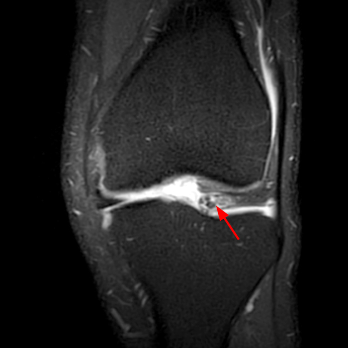
Meniscal Tear Patterns Radsource

How To Read Knee Mri Of Acl Tear Anterior Cruciate Ligament Injury Colorado Knee Surgeon Youtube Cruciate Ligament Knee Mri Acl Tear
Pin By Fateme On Bone And Joints Mri Brain Knee Mri Bone And Joint

Knee Anatomy Mri Knee Coronal Anatomy Free Cross Sectional Anatomy Mri Knee Mri Radiology

The Double Pcl Sign Appears On Sagittal Mri Images Of The Knee When A Bucket Handle Tear Of A Meniscus Medial Radiology Bone And Joint Radiology Technologist

Medpix Case 1 Right Acl Tear 2 Grade Ii Right Mcl Tear 3 Right Medial Meniscus Posterior Horn Tear
Https Encrypted Tbn0 Gstatic Com Images Q Tbn 3aand9gcsjqqaezr2k2wmmec3 Ivdizfnf8xzh4igpue1pn8gitv0lwel7 Usqp Cau
The Knee Musculoskeletal Key

Case Study Management Of Medial Meniscal Tear And Patellar Osteochondral Damage In A 60 Year Old Female

Torn Meniscus Symptoms And Causes Meniscus Tear Knee Mri Common Knee Injuries

Pin On Bone And Joints

Posterolateral Corner Injury Of The Knee Radiology Reference Article Radiopaedia Org
Https Pubs Rsna Org Doi Pdf 10 1148 Radiographics 22 4 G02jl03775

Pin By Dr Abuaiad On Bone And Joints Bone And Joint Knee Mri Mri

3m Splinting Guide Orthopedic Nursing Pediatric Nursing Pediatric Urgent Care

Pin By Juliann Marie On Msk Radiology Knee Mri Mri Radiology

Discoid Lateral Meniscus There Is A Discoid Lateral Meniscus Without Evidence Of A Lateral Meniscal Tear Knee Mri Radiology Medical Ultrasound

Can Knee Braces Help You Recover From A Torn Meniscus Injury Mcdavid

A Color Mri Medical Images Showing The Knee Joint Meniscus In Blue A Novel Advanced Visual Tool To See And Understand Anatomy Di Knee Joint Knee Mri Anatomy

Meniscal Flipped Fragment Radiology Case Radiopaedia Org

Pin By Dr Abuaiad On Bone And Joints Anatomy Of The Knee Bone And Joint Knee Mri

Cross Sectional Anatomy Of The Knee Based On Mri Articular Surface Of Patella And Femur Condyle Epicondyle And Muscles Popliteus Sar Knee Mri Mri Anatomy

Msk Radiology Panosundaki Pin

Mendmyknee Com Symptoms Of A Meniscus Injury Meniscus Tear Knee Surgery Recovery Knee Surgery

Mri Knee Anatomy Knee Sagittal Anatomy Free Cross Sectional Anatomy Mri Knee Mri Anatomy

Celery Stalk Sign Describes The Appearance Of An Anterior Cruciate Ligament That Has Undergone Mucoid Degeneration A Co Radiology Imaging Radiology Knee Mri

An Unusual Presentation Of Synovial Chondromatosis Of The Knee In A 10 Year Old Girl Sciencedirect

Sports Related Injuries Of The Knee An Approach To Mri Interpretation Stress Fractures

Pdf Surgical Management Of Degenerative Meniscus Lesions The 2016 Esska Meniscus Consensus

Epos

Shoulder Anatomy Mri Shoulder Axial Anatomy Free Cross Sectional Anatomy Shoulder Anatomy Axial Mri
1
Source : pinterest.com
